
Tackling Tough Anatomy with Modern Endodontic Technology
 One of the most rewarding facets of endodontics is seeing evidence of healing and successful treatment upon reevaluation at our follow-up visits. Although relieving the patient’s pain and eliminating swelling is of primary concern, it is always a great feeling to have patients witness the healing process through radiographic follow-up.
Explanation of osseous fill after adequate treatment reinforces the need for root canal treatment, and provides evidence that the treatment has eliminated the source of infection.
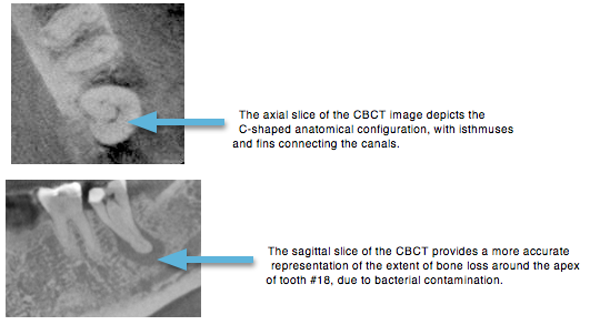
Understanding the anatomy of the root canal system is essential to achieve a positive outcome. This case is a perfect example of complex anatomy. The anatomical configuration was confirmed with the use of Cone Beam Computed Tomography (CBCT), which provides the best assessment of the tooth morphology and surrounding structures, via 3D analysis. The CBCT demonstrated a C-shaped configuration, which has been shown in endodontic literature to be difficult to adequately disinfect, due to the presence of isthmuses and fins.
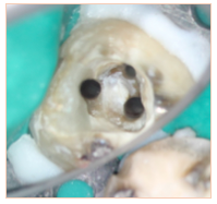
This clinical photo of the canals demonstrates the C-shaped nature of the anatomy. To aid in the disinfection process, Calcium hydroxide (Ca(OH)2) was placed as an inter appointment medicament for approximately 10 days.
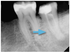
The radiopaque paste in the canal is the Ca(OH)2, which acts as a local antibiotic inside the tooth, to remove the source of the infection, bacteria.
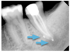
The patient returned for completion of the root canal 10 days after treatment was initiated. The post-op radiograph reveals sealer puffs along the lateral aspect of the root, which indicates a lateral canal was also filled. The core-buildup was placed and the patient was referred back to the referring general dentist for a full coverage crown.
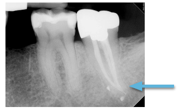
The patient returned for one year follow-up without complaint of pain/swelling. A crown was placed by the general dentist soon after completion of the root canal treatment. Complete healing is evident, with osseous fill and the re-establishment of a normal PDL space and lamina dura.
Thanks for visiting us at Ocean MicroEndodontics of San Diego! Visit back often for more great blog posts regarding our cases and other happenings here are our practice.
For more about C-shaped anatomy, check out the following review: The C-shaped Root Canal Configuration.